correct answer
e Mesoportal shunt (Rex shunt)
Portal hypertension in children can arise due to various factors, which generally fall into prehepatic, intrahepatic, and posthepatic categories. The following table summarizes the causes of portal hypertension in children.
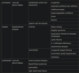
This patient’s history, physical exam, and laboratory findings are consistent with a prehepatic cause, namely extrahepatic portal vein thrombosis. This patient has preserved hepatic function with splenomegaly and his labs are consistent with hypersplenism. Causes of portal vein thrombosis include use of umbilical vein catheters, hypercoagulable conditions, and associated congenital anomalies including a greater than 10% incidence of cardiac anomalies requiring surgical repair.
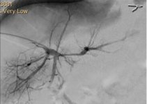
Imaging is critical for preoperative planning. Along with ultrasound (US) with Doppler, cross-sectional imaging (with venous phases) should be obtained. Intrahepatic portal vein branches are generally not well visualized even with cross sectional imaging. Portal venogram is necessary to clearly map the anatomy of the portal system. The image demonstrates a patent right and left branches of the portal vein.
A meso-portal (Rex) shunt is indicated for this patient with extrahepatic portal vein occlusion and a patent portal system. Continuing medical management only will lead to further bleeding episodes. TIPS is usually used as a bridge to transplant and data in young children are limited. Evaluation for liver transplant at this time is not warranted given the patient’s preserved liver function.
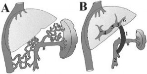
Schematic view of the portal venous system: (A) in patients with portal vein thrombosis and cavernomatous transformation and (B) after direct liver reperfusion by mesdportal bypass.