

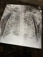
A 4-year-old was admitted to the PICU in ARDS. Ventilator settings include 100% FiO2, PRVC with TV of 5.5 ml/kg, RR 28, PEEP 12mmHg. Arterial blood gas showed: pH 7.42, pCO2 53, pO2 75, HCO3 34, base excess 8. You were called to help manage this finding on her CXR. It was interpreted as “significant pneumomediastinum, no pneumothorax, and significant subcutaneous emphysema."
On examination, patient has crepitus to the forehead and upper extremities to the wrist, back to iliac crests.
Examination did not show any pharyngeal injury. Esophagoscopy and bronchoscopy demonstrated mucosal integrity. The next best step is
a jet ventilation or high frequency oscillatory ventilation
b multiple incisions in areas of crepitus
c mediastinal tube placement
d bilateral chest tubes
e bilateral infraclavicular incisions
On examination, patient has crepitus to the forehead and upper extremities to the wrist, back to iliac crests.
Examination did not show any pharyngeal injury. Esophagoscopy and bronchoscopy demonstrated mucosal integrity. The next best step is
a jet ventilation or high frequency oscillatory ventilation
b multiple incisions in areas of crepitus
c mediastinal tube placement
d bilateral chest tubes
e bilateral infraclavicular incisions


