• Welcome to Pediatric Surgery Club! If this is your first visit, please sign up to get the best experience sign up!

weekly question 23/8/2026

.US site, click here!

Admin

Administrator
Staff member
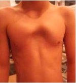
A 14-year-old boy presents with a chest wall deformity, pictured below. His mother says his chest looked abnormal at 7-years-old. He has chest pain and exercise intolerance. He and his parents want to improve the appearance of his chest. After chest CT and echocardiogram, which of the following is the most appropriate management?
repview (6).jpeg

a Trial of bracing

b Hybrid Ravitch/Nuss procedure

c Observation for two years

d Trial of combined bracing and vacuum bell

e Sternal rotation
 
correct answer
b Hybrid Ravitch/Nuss procedure

This patient has pectus arcuatum. The pathoanatomy of this chest wall deformity involves premature obliteration of the sternal sutures or congenital non-segmentation of the sternum, which results in angulation of the bone. This is distinct from both pectus carinatum and excavatum, where aberrant cartilage growth leads to an increase or decrease, respectively, in the AP diameter of the chest. Although similar in appearance, pectus arcuatum also differs from "mixed" pectus lesions, which result from abnormal cartilage development that leads to simultaneous protrusion and depression of the sternum. Bracing and/or vacuum bell therapy is not effective in pectus arcuatum. A hybrid surgical approach, consisting of a transverse incision, subperichondrial resections of upper costal cartilages, a sternal osteotomy and a low-placed Nuss bar, is a good option for definitive correction with minimal morbidity and high patient satisfaction. Another procedure with a vertical incision, subperichondrial cartilage resections, one or more sternal osteotomies and sternal plating is also effective. Sternal rotation would not be helpful with this pathology.
 
Back
Top