• Welcome to Pediatric Surgery Club! If this is your first visit, please sign up to get the best experience sign up!

weekly question 2/8/2026

.US site, click here!

Admin

Administrator
Staff member
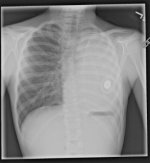
A 10-year-old child presents 3 months after undergoing a left pneumonectomy for a severe crush injury to the chest. They complain of stridor and shortness of breath with mild exercise and exertion. Their CXR and chest CT are shown below. What is the best next step for management of this patient’s symptoms?
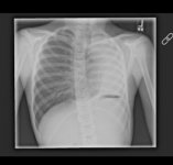
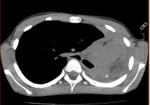
repview (3).jpegrepview (4).jpeg
a tracheal/bronchial resection with reanastomosis

b placement of intrathoracic prosthesis

c placement of endobronchial stent

d chemical blockage of phrenic nerve

e aortopexy
 
correct answer
b placement of intrathoracic prosthesis

Post-pneumonectomy syndrome (PPS) is rare and caused by dynamic airway obstruction due to mediastinal rotation following pneumonectomy. It is more common in children than adults due to an elastic mediastinum and also occurs more frequently after a right pneumonectomy. Following a pneumonectomy, the mediastinum can shift excessively towards the pneumonectomy space with associated rotation of great vessels and surrounding structures ultimately leading to extrinsic compression of the trachea or bronchus against the descending thoracic aorta and/or spine. The remaining lung herniates to the other side and becomes hyper-inflated. Patients with PPS may present with progressive dyspnea on exertion and stridor leading to reduced exercise capacity, tracheomalacia or respiratory failure requiring respiratory support. Diagnostic work-up can include CXR and chest CT as well as bronchoscopy.

Surgical correction of PPS should focus on restoring the mediastinum to its normal anatomic relationships. Various procedures have been tried with different degrees of long-term success. These include placement of endobronchial stents, crush or chemical blockage of the phrenic nerve to raise the hemidiaphragm, suture fixation of the aorta to the sternum, and tracheal/bronchial resection with reanastomosis anterior to the aortic arch.

Intrathoracic prosthesis placement with either a fixed-volume prosthesis and/or saline-filled tissue prosthesis is the preferred initial management for PPS. Tissue expanders with a remote port offer a safe way to adjust the intrathoracic volume, especially as a child grows. The main risk of implants are under/over filling and leakage of the expander. Recurrence of symptoms requires evaluation for the possibility of implant failure. In a recent review of adults with PPS, any type of surgical management without a prosthetic implant had a 100% incidence of recurrence.

repview(5).jpeg.029c8bf74a0ed8a617642b9dcd492b34.jpeg
Chest Radiograph with Prosthesis
 
Back
Top