Correct answer
d right thoracotomy.
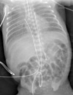
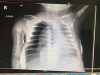
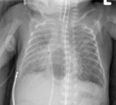
Given the history of extreme prematurity and difficult intubation this patient likely sustained a tracheal injury. The first radiograph shows the endotracheal tube (ETT) to be in the right mainstem bronchus. The child develops an additional pneumothorax. A small posterior pneumomediastinum is seen on the second film. Despite changes in the ventilation strategy and placement of a chest tube, the child continues to have a large air leak. While hyaline membrane disease, surfactant deficiency, is in the differential, the large air leak and difficulty intubating the child raises a concern for tracheobronchial injury. Further observation is not appropriate given the progression of the bronchial air leak.
Iatrogenic tracheal injuries can occur with difficult and traumatic intubations. A common presentation is posterior pneumomediastinum as seen in the initial radiographs above. Management is largely based on the condition and size of the patient. Intubation while under general anesthesia prior to surgery is a common scenario for iatrogenic tracheal injuries. In such instances when tracheal injuries are suspected, bronchoscopy should be performed to determine the location and extent of the injury. In stable patients, small injuries can be managed with extubation and close observation. If the injury is in the proximal trachea, the ETT can be positioned to cover the injury. Repeat bronchoscopy in one week will often demonstrate complete healing of the injury and the patient can be safely extubated. In some cases, bronchoscopy may not be possible due to the small patient size.
Observation is the first step in management of premature neonates with suspected tracheal injury. If tolerated, patients should be extubated to avoid positive pressure ventilation. If unable to extubate, then ventilation with the high-frequency oscillator should be attempted. In this case, the pneumomediastinum significantly increased despite conservative management including ventilation changes and surgical intervention is required. The best approach for tracheal injuries is via right thoracotomy as the entire trachea can be exposed. Injuries can often be primarily repaired and reinforced with a pleural flap.
In premature infants, single lung ventilation via right mainstem intubation is typically not tolerated and thus would not be a viable option in this patient. A second chest tube could be considered; however, given the large air leak it is unlikely that this will obtain pleural apposition to seal the leak. Exposure via median sternotomy will not be adequate for repair.
Although tracheal injuries are rare and only case reports are available, pediatric surgeons should be familiar with the management of this condition.